Kyoto: Researchers from Kyoto University, Japan have created a new device that can culture cancerous cells in vitro in a three-dimensional fashion that better mimics the conditions present in the human body. Touted as ‘tumour-on-a-chip’, this piece of equipment opens up new possibilities for testing several potential cancer-fighting drugs with greater accuracy and ease.
The details of this new invention were revealed in the paper “Vascularized cancer on a chip: The effect of perfusion on growth and drug delivery of tumour spheroid” in the journal Biomaterials.
The path to drug discovery is never easy. Scientists and clinicians can go through tens-of-thousands of potential compounds for years to find a handful of viable candidates, only for them to fail at the clinical level.
“Potential compounds are tested using animal models and cells cultured in a dish. However, those results frequently do not transfer over to human biology,” explains first author Yuji Nashimoto formally of the Graduate School of Engineering, and now at Tohoku University. “Furthermore, cells on a dish lack the three-dimensional structure and blood vessels, or vasculature that keeps it alive. So, we came up with a plan to construct a device that solves these issues.”
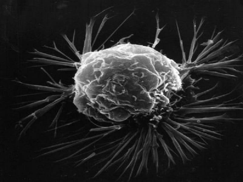
The device, reported in the journal Biomaterials, is the size of a coin with a 1 mm well at the center. This well is flanked by a series of 100 mm ‘microposts’. The idea is that a three-dimensional culture of tumor cells is placed in the middle well, and then cells that constrict blood vessels are places along the micro posts. Over a few days, the vessels grow and attach to the culture.
“This ‘perfusable vasculature’ allows us to administer nutrients and drugs into the system to mimic the environment in the body,” continues Nashimoto. “This allows us to have a clearer picture of the effectiveness of cancer-treating compounds.”
This perfusion did significantly keep the tumor cells healthy by keeping cell proliferation up and cell death down. A drug assay was then performed with the team administrating an anti-tumor drug at low doses. Interestingly, the drug was more effective under static conditions compared to when nutrients were flowing through the tumor cells.
In contrast, the drug’s effects became more potent when the flow was turned on and the dosage was increased. Ryuji Yokokawa, who leads the team, explains that the unexpected results prove that we need to consider the balance between the proliferation of tumor cells and the efficacy of the drug under flow conditions.
“We hypothesize that at low doses the benefit of the nutrient flow outweighs the effect of the anti-tumor drug. It proves the importance of blood flow in the vasculature when screening for drugs.”
He concludes, “Due to its size and utility, we hope the new device can expedite the tests on the countless number of potential new drugs. While many questions remain, we are happy to have developed this device and have shown that three-dimensional perfused cell culture is vital for the next step in drug discovery.”